Cellular and Physics Microbiology of Infection
INSERM U1019 – CNRS UMR9017 – Lille University – CHU Lille – Institut Pasteur de Lille

Presentation
The Cellular Microbiology and Physics of Infection team is developing a multidisciplinary approach to study the influences of membrane mechanics on the cellular response to infection. Our studies use methods borrowed from cellular and molecular biology, imaging including super-resolution and correlative photonic/electronic microscopy, biophysics of molecular interactions and elasticity of cells and tissues. The question posed is how membrane interactions can modulate the activation of signaling cascades resulting from infection. The signalling pathway studied is autophagy rather in its usurpation by the microorganism. We focus our studies on the early stages of bacterial infection related to different diseases (shigellosis, listeriosis, yersiniosis, meningococcal meningitis, nosocomial), intoxinations (Anthrax) and viral infections (Norovirus) and also for the importance of the microbiota (acid lactic acid bacteria). We have chosen different scales of intermolecular interactions on membrane models that support intercellular and even tissue interactions.
Highlights
Autophagy is a ubiquitous process of eliminating sub-compartments within cells for recycling (Y Ohsumi Nobel Prize 2016) and can be subverted by pathogens. Our team has uncovered fundamental processes in microbiology such as the involvement of molecular complexes linked to autophagy in the elimination of danger signals in shigellosis (bacillary dysentery), the usurpation of autophagic membranes in cases of internalization of Y pseudotuberculosis (responsible for entheropathies) or for the repair of the vacuolar compartment of Coxiella burnetii (responsible of the Q fever zoonosis). Some of our discoveries have been extended to other pathogens (bacteria and viruses) or even demonstrated as fundamental cellular processes.
Our research depends on the development of nanotechnologies such as fluorescence nanoscopy (Nobel Prize 2014 for E Betzig, SW Hell & WE Moerner) or force spectroscopy using near-field microscopy (Nobel Prize 1986 for G Binnig & H Rohrer). For this, we have a unique park of atomic force microscopes, some of which are coupled with super-resolution microscopes in biophotonics (financed thanks to the ANR Equipex ImagInEx BioMed / FEDER future investment plan program coordinated by F Lafont). We have developed innovative and original approaches such as hardness tomography, which allows us to measure the elasticity of intracellular compartments, correlative microscopy combining the three microscopic methods of atomic force, electron and photonics or the standardization of elasticity measurements on cells (European consortium COST AFM4nanomed&bio) for which we are internationally recognized specialists.
In addition, thanks to these projects of excellence we have multiple collaborations that have enabled us to provide essential data among others for the identification of the meningococcus receptor (S Bourdoulous I Cochin Paris), the characterization of transient cell perforations resulting from anthrax intoxination (E Lemichez, I Pasteur Paris).
Transversal projects
 Usurpation of autophagy pathways by pathogens
We want to characterize molecular complexes involving proteins linked to the autophagy machinery playing a role in the interactions between host cells and pathogenic bacteria using different infection strategies (ANR StopBugEntry). The underlying idea being that by inhibiting these mechanisms, the infectious process would be prevented. In addition, we want to uncover previously ignored or underestimated phenomena such as the extinction of signaling associated with pathogen internalization or breakage processes – repair of vacuolar compartments (Coop Inter CNRS, ANR mitopatho).
Determination of (nano-)mechanobiological properties on cells and tissues
Thanks to our innovative technological developments notably related to membrane elasticity measurements and correlative approaches combining several types of microscopy we can establish standards and new approaches (EU-COST-AFM4NanoMed&Bio, EU-ITN-Phys2bioMed) to help diagnose certain diseases beyond infection such as cancer. These developments are made available to the scientific and technological communities (EU-COST Tree-Mobieu) and allow conceptual advances capable of explaining transient cell opening mechanisms (ANR TransEndothelia tunnel). This process could play an important role in transcytosis phenomena.
Structure-function characterization of infectious processes
We develop multi-scale approaches to better understand the membrane phenomena involved in host-pathogen interactions. We adopt a reductionist methodology with the nano-characterization of the behavior of proteins inserted in suspended lipid bilayers (ISITE STAND). In addition, we characterize the structure of pathogen membranes as a function of conditions by considering lipids (MtbLipVir RNA) or protein self-assembly (Funhydro RNA). Finally, we consider pathogens and the host cell interacting to understand the mechanobiological processes involved such as during the retraction of pili during meningococcal adhesion (RNA Retractopath).
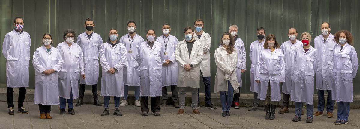
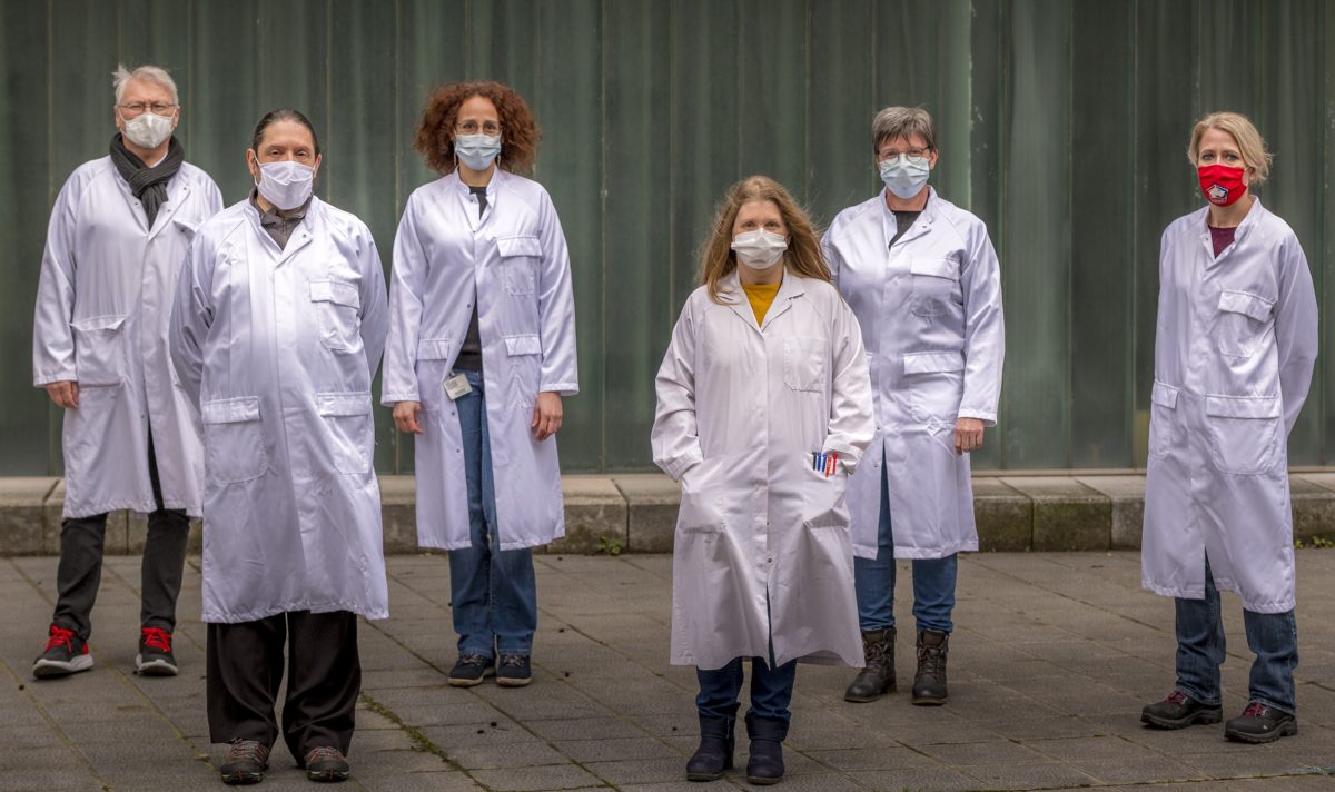
Members
Frank LAFONT
DR1 CNRS, group leader
ORCID number : 0000-0001-8668-2580
Lorena REDONDO-MORATA
CRCN Inserm
ORCID number : 0000-0002-6254-8261
Fabio PERISSINOTTO
Post-doc, Inserm
ORCID number : 0000-0002-8123-0793
Maria-Luisa MARINI
Doc EU-ITN
Vincent DUPRES
MCU, Univ Lille
ORCID number : 0000-0002-1970-3633
Joëlle WAREIN
Tech IPL
Javier LOPEZ
Doc EU-ITN
Ghaffar MUHARRAM
MCU, Univ Lille
ORCID number : 0000-0003-4309-8450
Ahmad MELHEM
IE CNRS
Emmrich WAKEFORD
Master, Univ Lille
Tristan BERTRAND
Master, Univ Lille
Amaury WUILBAUT
Licence 3, Univ Lille
Catherine DANIEL
CR IPL
ORCID number : 0000-0002-5299-2799
Sabine POIRET
Tech IPL
Simon LAMBERT
Master, Univ Lille
Nicolas BAROIS
IR Inserm
Sébastien JANEL
IR CNRS
ORCID number : 0000-0001-6736-3162
Elisabeth WERKMEISTER
IR CNRS
ORCID number : 0000-0002-6253-7503
Sébastien LECOMTE
BTS, V. Labbé
Publications
Janel S , Popoff M , Barois N , Werkmeister E , Divoux S , Perez F , Lafont F . (2019)
Stiffness tomography of eukaryotic intracellular compartments by atomic force microscopy.
Nanoscale. May 30;11(21):10320-10328.
Dujardin A, De Wolf P, Lafont F, Dupres V. (2019)
Automated multi-sample acquisition and analysis using atomic force microscopy for biomedical applications.
PLoS One. Mar 15;14(3):e0213853. doi: 10.1371/journal.pone.0213853.
Ciczora Y, Janel S, Soyer M, Popoff M, Werkmeister E, Lafont F. (2019)
Blocking bacterial entry at the adhesion step reveals dynamic recruitment of membrane and cytosolic probes.
Biol Cell. Mar;111(3):67-77. doi: 10.1111/boc.201800070.
Denis K, Le Bris M, le Guennec L, Barnier J-P, Faure C Gouge A, Bouzinba-Ségard H, Jamet A, Euphrasie D, Durel B, Barois N, Pelissier P, C. Morand P, Coureuil M, Lafont F, Join-Lambert O, Nassif X, Bourdoulous S. (2019)
Targeting Type IV pili as an antivirulence strategy with phenothiazines protects against Neisseria meningiditis infection.
Nat. Microbiol. 4:972-984.
Radulovic M, Schink KO, Wenzel EM, Nähse V, Bongiovanni A, Lafont F, Stenmark H; (2018).
ESCRT-mediated lysosome repair precedes lysophagy and promotes cell survival.
EMBO J. Nov 2;37(21):e99753.
Keywords
Autophagy ; Host-pathogen interaction ; Bacteria ; Toxin ; Norovirus ; Lactic acid bacteria ; Mecanobiology ; Correlative microscopy ; Atomic Force Microscopy